The scientific objectives
The LOOP project aims to bring together the University of Lille’s theoretical, technological and clinical forces in neurotechnology, more specifically those involved in the different stages of designing, testing and validating devices that can mimic and restore brain physiology, with the ultimate goal of relieving severe psychiatric symptoms. The CDP will focus on the multidisciplinary field of closed-loop systems in neuroscience, which can be understood as automated control systems regulated by feedback. The project will mobilize several Lille partners highly involved in the NeurotechEU European alliance in order to take advantage of all the necessary expertise from physics, materials, microelectronics and nanotechnology, computer science and neuromorphic computing, signal processing, cognitive neuroscience, clinical research, ethics and law.

The image displays the logos of all the scientific partnairs involved in the LOOP project
- Lille Neuroscience & Cognition
- IEMN (Institut d’Electronique, Microélectronique et Nanotechnologie)
- CRIStAL (Centre de Recherche en Informatique, Signal et Automatique de Lille)
- SCALab (Sciences Cognitives et Sciences Affectives)
- CRDP (Centre de Recherche Droits et Perspectives du droit)
- CHU Lille
- Université de Lille
- UFR3S
- INM (Institut de Neuromodulation)
- IRCICA (Institut de Recherche sur les Composants logiciels et matériels pour l'Information et la Communication Avancée)
- NeurotechEU: the European University of Brain and Technology
This CDP will build on clear evidence from the fields of neuroprosthetics and brain-machine interfaces (BMI), in which it has been demonstrated that closed-loop paradigms can be highly successful, for example in the use of neurofeedback in cognitive rehabilitation.
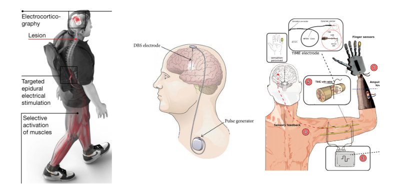
1. Left Section: Electrocorticography and Targeted Electrical Stimulation
-
Main Elements:
- Electrocorticography (ECoG): Electrodes are placed directly on the cerebral cortex to record electrical activity.
- Lesion: An area of the brain is marked as damaged, likely indicating a region affected by stroke or injury.
- Targeted Epidural Electrical Stimulation: Electrodes are used to stimulate the spinal cord specifically.
- Selective Activation of Muscles: Electrical stimulation enables targeted activation of leg muscles, aiding in walking.
-
Description:
This section demonstrates how recording brain activity via electrocorticography can be used to selectively activate leg muscles, assisting in the rehabilitation of walking for patients with brain lesions.
2. Middle Section: Deep Brain Stimulation (DBS)
-
Main Elements:
- DBS Electrode: An electrode for deep brain stimulation is implanted in the brain.
- Pulse Generator: A device, typically implanted in the chest, generates electrical pulses sent to the DBS electrode.
-
Description:
This illustration shows the principle of deep brain stimulation (DBS), used to treat various neurological disorders by sending electrical impulses to specific brain areas.
3. Right Section: Sensory Neurofeedback and Transcranial Stimulation
-
Main Elements:
- Finger and Hand Sensors: Sensors on a prosthetic hand record movements and send sensory information.
- TIME Electrodes: Transcranial electrodes stimulate the brain to provide sensory feedback.
- Sensory Neurofeedback: The system uses sensory signals to provide feedback to the brain, enhancing perception and movement control.
- Transcranial Stimulation: An electrode stimulates the brain non-invasively.
-
Description:
This section describes a sensory neurofeedback system where sensors on a prosthetic hand record movements. This information is used to stimulate the brain via transcranial electrodes, allowing sensory feedback and improving control of prosthetics.
Towards innovative closed-loop therapies for schizophrenia
To illustrate the innovative potential of closed-loop therapies in psychiatric disorders, the project will implement an interdisciplinary strategy applied to the development of a mechanism-based therapy for drug-resistant hallucinations in patients with schizophrenia. The project will be structured around three main research pillars to unlock the current bottlenecks preventing the deployment of closed-loop therapies, alongside two horizontal, cross-cutting strands: ethics and law on one side, and education on the other.
- The first vertical research pillar will bring together scientists to work on the algorithmic level of closed-loop systems (online signal processing, machine learning).
- The second pillar will unite research groups around the design of a complete and non-invasive solution to address the question of refractory hallucinations, and study how closed-loop interaction schemes can be beneficial for highly impaired psychiatric patients.
- The third research pillar will be devoted to the design of the sensors themselves and translational research with animal models, in order to enhance the quality of the recordings and pave the way for future invasive miniaturized solutions.
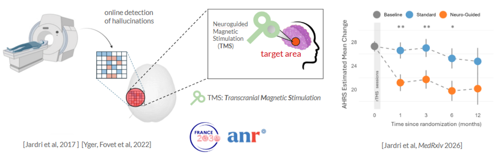
The image illustrates an innovative approach that combines medical imaging and transcranial magnetic stimulation (TMS) to target hallucinations.
From left to right:
1. MRI (Magnetic Resonance Imaging) machine:
- On the left, an illustration of an MRI machine shows how brain images are acquired. These images allow specific areas of the brain to be detected.
2. Real-time detection of hallucinations:
- A colored grid (with red and blue squares) symbolizes the analysis of brain data to detect hallucinations in real time.
3. Targeting the brain area:
- A stylized human head shows a targeted brain area (in red) where hallucinations are detected.
4. Neuroguided Magnetic Stimulation (TMS):
- A TMS coil is positioned on the head to stimulate the targeted brain area. TMS (Transcranial Magnetic Stimulation) is used to modulate activity in this area.
Results graph:
On the right, a graph shows the change in the AHRS (Auditory Hallucination Rating Scale) Estimated Mean Change over a 12-month period following randomization. Three curves are shown:
- Gray (Baseline)
- Blue (Standard)
- Orange (Neuro-Guided)
- The results indicate a more pronounced reduction in hallucinations with the neuro-guided method (orange) compared to the standard method (blue).
References:
The image cites scientific studies, including [Jardri et al., 2017], [Yger, Fovet et al., 2022], and [Jardri et al., MedRxiv 2026], as well as logos for research project funding (France 2030, ANR).